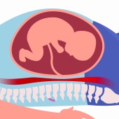
Chorionic villus sampling (CVS) is a prenatal test that involves taking a sample of tissue from the placenta to test for chromosomal abnormalities and certain other genetic problems. The placenta is a structure in the uterus that provides blood and nutrients from the mother to the fetus,
The chorionic villi are tiny projections of placental tissue that look like fingers and contain the same genetic material as the fetus. Testing may be available for other genetic defects and disorders depending on the family history and availability of lab testing at the time of the procedure.
CVS is usually performed between the 10th and 12th weeks of pregnancy. Unlike amniocentesis (another type of prenatal test), CVS does not provide information on neural tube defects, such as spina bifida. For this reason, women who undergo CVS also need a follow-up blood test between 16 to 18 weeks of their pregnancy to screen for neural tube defects.
There are two types of CVS procedures:
Transcervical: In this procedure, a catheter is inserted through the cervix into the placenta to obtain the tissue sample.
Transabdominal: In this procedure, a needle is inserted through the abdomen and uterus into the placenta to obtain the tissue sample.
- 6371 views

