A racheoesophageal fistula is a birth defect in which the esophagus has an abnormal connection to the trachea. The esophagus is the tube that food passes through from the mouth to the stomach, and the trachea is the windpipe that air passes through from the mouth and nose to the lungs.
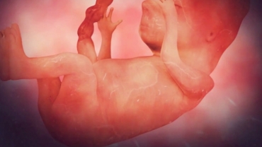
The trachea forms during the sixth week of pregnancy. Here, you can see the tube that will eventually form all the organs of the digestive system. The trachea and lungs grow from the part of the digestive tube that will eventually become the esophagus. For unknown reasons, the esophagus and trachea may grow and separate abnormally during this time.
The esophagus may end in a blind pouch with missing gaps and be abnormally narrow. This absence or narrowing of a natural body passageway is called atresia. The esophagus and trachea may also have an abnormal connection called a tracheoesophageal fistula. Here, we see the most common type of tracheoesophageal fistula in a newborn infant.
The upper esophagus ends in a blind pouch, and the lower esophagus connects to the trachea. This is a serious problem because stomach contents can travel up the esophagus and pass through the fistula into the trachea and lungs. The fistula can also cause difficulty breathing for the newborn since air can now bypass the lungs and enter the stomach.
Before the tracheoesophageal fistula repair procedure, an intravenous line will be started. The baby may be given antibiotics through the IV to decrease the chance of infection. The baby will be given general anesthesia which will put the baby to sleep for the entire operation. A breathing tube will be inserted through the mouth and down the throat to help the baby breathe during the operation.
The surgeon will make an incision in the baby’s chest, usually on the right side. Through the incision, the surgeon will gently move the lungs aside to view the trachea and the esophagus. After identifying the tracheoesophageal fistula, the surgeon will slowly close the fistula’s connection with the trachea with sutures, then cut the connection away from the trachea. The fistula’s connection with the esophagus will also be cut, and the fistula will be removed. Next, the surgeon will make an incision at the end of the upper esophagus to open it. Then, the upper and lower esophagus will be connected with sutures.
Finally, the surgeon will insert a surgical drain in the chest and close the incision with sutures. After the procedure, the baby will continue to use the breathing tube until they’ve healed enough to breathe on their own. The baby will be taken to the neonatal intensive care unit for monitoring. Pain medication will be given. The baby may continue to receive antibiotics through the IV. Babies are released from the hospital when they’re able to eat enough to maintain their weight, which may be after two weeks or longer.
- 18 views

